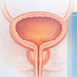
Bladder Volume Calculator and Report Generator
Related Calculators
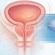
Prostate volume estimation with PSA density calculation using imaging measurements and serum PSA levels.

Splenic volume and index estimation using imaging measurements with reference to normal size ranges.
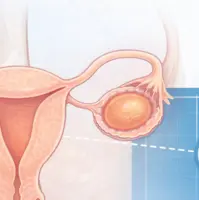
Gonadal volume estimation using imaging measurements for evaluation of size, development, and pathology.
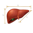
Liver volume estimation using imaging measurements with comparison to expected normal values.
More about the Bladder Volume Calculator
Bladder volume calculation in daily clinical imaging
The Bladder Volume Calculator should be understood within the broader context of bladder volume estimation, rather than as an isolated tool. In radiology and related clinical settings, bladder volume estimation supports assessment of urinary retention, post void residual measurement, perioperative monitoring, and radiotherapy preparation. Ultrasound is widely used because it is noninvasive, repeatable, and readily available. The reported value reflects a calculated estimate based on measured dimensions and an assumed geometric model, not a direct measurement of bladder contents.[1][2][3]
Interpretation depends on how the measurements were obtained and how closely the selected model reflects the actual bladder contour. For radiologists, the numerical result is best reviewed together with the images rather than treated as a standalone finding.
Clinical role of bladder volume ultrasound
Bladder ultrasound for volume estimation is commonly used when catheterization is undesirable or when repeated measurements are needed. Typical indications include evaluation of urinary retention, post void residual assessment, inpatient monitoring, and preparation for pelvic radiotherapy where reproducible bladder filling is important.[2]
Small differences in caliper placement can lead to noticeable differences in calculated volume because the final value is derived from multiple orthogonal measurements rather than a single dimension.
Measurement principles and bladder volume formulas
Most methods of bladder volume calculation rely on three orthogonal measurements, length, width, and height, multiplied by a correction coefficient. This is a practical geometric approximation rather than a direct anatomic measurement. The bladder does not maintain a fixed shape, and its contour changes with filling, pelvic anatomy, and surrounding structures.[4]
Standard ultrasound measurement technique
When measuring bladder dimensions on ultrasound:
- Length is measured in the sagittal plane from base to dome.
- Height or depth is the maximal anteroposterior dimension, usually in the sagittal plane.
- Width is measured in the transverse plane.
Accuracy depends on identifying the true maximal dimension in each plane. Off-axis measurements, incomplete visualization of the bladder wall, and suboptimal acoustic windows can all influence the estimate.
Different geometric models use different coefficients to generate an estimated bladder volume. In this calculator, the selected shape determines which coefficient is applied to the measured dimensions. These coefficients reflect different modeling assumptions and should be interpreted as shape-based approximations rather than interchangeable universal standards.[3][4][5]
| Modeled bladder shape | Constant | Formula | Interpretive context |
|---|---|---|---|
| Unknown shape | 0.72 | 0.72 × L × W × H | Reported as a general approximation when contour does not clearly fit a specific model. |
| Elongated or cylindrical approximation | 0.81 | 0.81 × L × W × H | Research-based approximation that may better fit some distended or elongated contours. Not equivalent to the standard 0.52 ellipsoid formula. |
| Cuboid approximation | 0.89 | 0.89 × L × W × H | Assumes less tapering of the bladder wall. |
| Spherical approximation | 0.52 | 0.52 × L × W × H or 0.52 × D3 | May be used when a more rounded contour is present. |
| Triangular prism approximation | 0.66 | 0.66 × L × W × H | Proposed for wedge-shaped configurations. |
These values are best understood as model-dependent coefficients drawn from the published literature. Their main value is that they show why the same measured dimensions can produce different estimated volumes when a different bladder shape is assumed.[3][4][5]
Measurement variability and image-based interpretation
Bladder shape changes with filling and surrounding anatomy. A partially filled bladder may appear ovoid, whereas a more distended bladder may become more rounded or assume a contour constrained by adjacent pelvic structures. Compression from nearby organs, postoperative change, trabeculation, or asymmetric filling can alter the relationship between measured dimensions and true intravesical volume.[4][5]
Variability arises from both technique and model selection. Relevant factors include caliper placement, image quality, patient body habitus, irregular bladder wall morphology, and differences in correction coefficients. In a foundational geometric modeling study, Bih and colleagues found that shape-specific correction coefficients reduced mean estimation error within that study population compared with use of a single global coefficient, illustrating why contour matters when interpreting calculated bladder volume.[4]
This is particularly relevant in post void residual measurement, where relatively small differences between measurements may fall within expected technical and modeling variability rather than representing a true physiologic change.
| Observed contour | Typical imaging appearance | Potential effect on estimation | Interpretive implication |
|---|---|---|---|
| Elongated or ovoid | Smooth contour with moderate filling | Often aligns reasonably well with elongated or ellipsoid-based estimation | Common in routine imaging. |
| Rounded or markedly distended | More symmetric contour with larger volume | May produce different estimates depending on the geometric model used | Relevant in retention assessment and reproducibility workflows.[2][5] |
| Flattened | Compression by adjacent structures | May reduce accuracy of simple geometric assumptions | Seen in postoperative settings or with mass effect. |
| Irregular | Trabeculated or distorted wall | Associated with greater variability | Requires careful image correlation. |
Bladder shape and consistency in radiotherapy
In radiation oncology, bladder filling influences reproducibility between treatment sessions. Huang and colleagues showed that bladder shape, including the relationship between height and width, affects setup consistency in prostate radiotherapy.[2]
From an imaging perspective, similar calculated volumes may not correspond to identical pelvic geometry if bladder shape differs between examinations. Reproducibility therefore depends on more than the numeric value alone.
Practical interpretation in clinical workflows
The Bladder Volume Calculator provides a structured way to organize measurements. The calculated value should be interpreted alongside image quality, observed bladder morphology, and the clinical scenario.
- Use consistent measurement planes.
- Confirm that each measurement represents the maximal dimension.
- Document whether the study is pre-void or post-void.
- Be cautious when comparing values derived from different models or devices.
- Correlate the numerical estimate with the observed contour when morphology is atypical.
In practice, bladder volume calculation is most useful when combined with careful image review and consistent technique.
Frequently Asked Questions (FAQs)
Why can the coefficient change based on bladder shape?
Different coefficients reflect different geometric assumptions. A bladder that appears rounded, elongated, flattened, or otherwise nonuniform may be approximated differently by a mathematical model, which can change the estimated volume even when the measured dimensions are the same.[3][4][5]
Why can two measurements of the same bladder produce different estimated volumes?
Differences may arise from caliper placement, selection of maximal dimensions, image quality, patient positioning, and the correction coefficient used. The same length, width, and height measurements can also yield different calculated volumes if different geometric assumptions are applied.
How should irregular or trabeculated bladders be interpreted?
Irregular wall contour can reduce the accuracy of simple geometric formulas because the bladder no longer closely resembles an idealized shape. In these cases, the calculated value should be interpreted more cautiously and correlated directly with the sonographic appearance.[4]
Why does shape matter in post void residual assessment?
In post void residual measurement, relatively small absolute volume differences can influence interpretation. Because shape and technique both affect the estimate, minor differences between serial measurements may reflect expected variability rather than a true physiologic change.
Why is bladder shape relevant in radiotherapy preparation?
For radiotherapy workflows, reproducibility of bladder filling between sessions is important. Shape can influence whether similar calculated volumes correspond to similar pelvic geometry, which is one reason the numeric estimate should not be considered in isolation.[2]
References
- Yuan J, Shen M, Zhang T, et al. A novel three-point localization method for bladder volume estimation. Sensors (Basel). 2024;24(6):1932. doi:10.3390/s24061932.
- Huang S, Li T, Guo Y, et al. Impact of bladder volume and bladder shape on radiotherapy consistency and treatment interruption in prostate cancer patients. J Appl Clin Med Phys. 2025;26(4):e70026. doi:10.1002/acm2.70026.
- Vinod NN, Nagle AS, Naimi HA, et al. Bladder volume correction factors measured with 3D ultrasound and BladderScan. Can J Urol. 2019;26(4):9829-9834.
- Bih LI, Ho CC, Tsai SJ, Lai YC, Chow W. Bladder shape impact on the accuracy of ultrasonic estimation of bladder volume. Arch Phys Med Rehabil. 1998;79(12):1553-1556. doi:10.1016/S0003-9993(98)90419-1.
- Chang WL, Lai SH, Cheng CF, Chiu V, Lin SK. Comparative effectiveness of two models of point-of-care ultrasound for detection of post-void residual urine during acute ischemic stroke: preliminary findings of real-world clinical application. Diagnostics (Basel). 2023;13(15):2599. doi:10.3390/diagnostics13152599.

PGY-5 Radiology and Nuclear Medicine Resident Physician
UT Southwestern Medical Center, USA
