PIRADS Calculator v. 2.1
Lesion-level chance of malignancy per PI‐RADS® v2.1 score, according to Oerther et al. ²:
- PI-RADS 1 – Very low (~2%)
- PI-RADS 2 – Low (~4%)
- PI-RADS 3 – Intermediate (~20%)
- PI-RADS 4 – High (~52%)
- PI-RADS 5 – Very high (~89%)
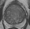
All images on this page are obtained from ACR's PI-RADS atlas.
References:
- Turkbey B, Rosenkrantz AB, Haider MA, et al. Prostate Imaging Reporting and Data System Version 2.1: 2019 Update of Prostate Imaging Reporting and Data System Version 2. Eur Urol. 2019;76(3):340-351. doi:10.1016/j.eururo.2019.02.033
- Oerther B, Engel H, Bamberg F, Sigle A, Gratzke C, Benndorf M. Cancer detection rates of the PI-RADSv2.1 assessment categories: systematic review and meta-analysis on lesion level and patient level. Prostate Cancer Prostatic Dis. 2022;25(2):256-263. doi:10.1038/s41391-021-00417-1
Related Calculators:

More about the PIRADS Calculator (v2.1):
The PIRADS calculator on our site is designed to assist clinicians and radiologists in applying the Prostate Imaging Reporting and Data System, version 2.1, in a clear, consistent, and efficient manner. This interactive tool aligns with the structured guidelines published by the American College of Radiology (ACR) to support the diagnosis, risk assessment, and management of prostate cancer through multiparametric MRI (mpMRI). While not officially endorsed by the ACR, our implementation reflects the framework's full diagnostic rigor and is ideal for clinical, educational, and decision-support applications.
What is PI-RADS v2.1?
PI-RADS v2.1 is a standardized reporting system that enhances the detection and risk stratification of clinically significant prostate cancer. It combines the interpretation of multiple imaging sequences, T2-weighted imaging (T2WI), diffusion-weighted imaging (DWI), and dynamic contrast-enhanced (DCE) imaging into a unified scoring system that ranges from PI-RADS 1 (very low suspicion) to PI-RADS 5 (very high suspicion).
- T2-Weighted Imaging (T2WI): Offers high-resolution images of the prostate anatomy, critical for identifying lesion location and morphology.
- Diffusion-Weighted Imaging (DWI): Evaluates cellular density, helping to distinguish between benign and malignant lesions.
- Dynamic Contrast-Enhanced Imaging (DCE): Captures vascular properties of lesions by analyzing enhancement kinetics post-gadolinium administration.
Why Use a PIRADS Calculator?
Manually assigning a PI-RADS score can be time-consuming and subject to interobserver variability. The PIRADS calculator simplifies this task by guiding users through a structured input process that mirrors the PI-RADS lexicon. It automatically integrates the findings from each sequence to calculate an overall score and risk level, reducing interpretation errors and enhancing efficiency in busy clinical settings.
Benefits of Using the PIRADS Calculator
Designed with practicality and precision in mind, this tool offers multiple advantages:
- Improved diagnostic consistency: Ensures uniform application of PI-RADS criteria across institutions and users.
- Time-saving automation: Reduces the cognitive and logistical load of manual scoring.
- User-friendly design: Suitable for both experienced radiologists and trainees, with intuitive input fields and automatic risk calculation.
- Evidence-based methodology: Follows the established clinical imaging criteria laid out in the PI-RADS v2.1 guidelines.
Clinical Application in Prostate Cancer Management
The PI-RADS scoring system plays a central role in guiding decisions about prostate biopsy, active surveillance, or advanced interventions. A score of PI-RADS 4 or 5 typically warrants further diagnostic action, while PI-RADS 1 or 2 may suggest watchful waiting or routine follow-up. By integrating this PIRADS calculator into workflow, clinicians can expedite the diagnostic process while maintaining a high standard of care. This aligns well with precision oncology practices and risk-based patient stratification.
Teaching and Research Applications
Beyond clinical use, this tool is also valuable for academic settings. It aids radiology residents, urology fellows, and medical students in mastering the interpretation of prostate MRI. Researchers involved in prostate imaging studies can also use the calculator to ensure consistency in lesion scoring and data collection. The tool’s structured approach supports reproducible research outcomes, especially in studies involving PI-RADS performance metrics.
Role in Multidisciplinary Prostate Cancer Care
Effective communication among radiologists, urologists, oncologists, and pathologists is essential in prostate cancer care. The standardized outputs generated by the PIRADS calculator promote this collaboration by presenting risk categories in a clear, universally accepted format. This allows different specialties to align on biopsy indications, treatment planning, and follow-up strategies.
PIRADS Calculator and PSA Density
Although PI-RADS focuses on MRI findings, combining it with other markers like PSA density improves diagnostic accuracy. Our prostate imaging tools, including PSA density calculators, can complement the PI-RADS score to refine risk assessment and biopsy decisions. The calculator becomes even more powerful when used alongside prostate volume estimates and serum PSA levels in an integrated diagnostic approach.
PI-RADS, Artificial Intelligence, and the Future
As artificial intelligence (AI) continues to shape medical imaging, PI-RADS-based tools are being integrated into machine learning models that automate lesion detection and scoring. Our calculator reflects these emerging trends by offering a digital, semi-automated interface that serves as a stepping stone toward more comprehensive AI-driven diagnostic systems. In this evolving landscape, tools like the PI-RADS calculator play a critical role in bridging human interpretation and algorithmic support.
Conclusion
The PIRADS calculator is a valuable resource for improving the clarity, consistency, and speed of prostate cancer imaging workflows. Whether you're a radiologist interpreting a complex mpMRI case, a urologist making a biopsy decision, or a student learning about prostate cancer diagnostics, this tool offers practical utility backed by evidence-based guidelines. By adhering to the principles of PI-RADS v2.1, it supports standardized reporting and enhances communication across the care team - ultimately contributing to better patient outcomes.
Frequently Asked Questions (FAQ)
- What does the PI-RADS calculator do?
It helps apply the PI-RADS v2.1 framework to prostate MRI findings in a structured way. The calculator guides users through interpreting T2-weighted, diffusion-weighted, and contrast-enhanced sequences to estimate a consistent PI-RADS category. - Who can use the PI-RADS calculator?
This tool is intended for healthcare professionals, radiology trainees, and researchers who interpret prostate MRI scans. It can also serve as a learning resource for those studying prostate imaging principles. - What is the purpose of PI-RADS?
PI-RADS (Prostate Imaging Reporting and Data System) provides a standardized method for describing prostate MRI findings and estimating the likelihood of clinically significant cancer. It helps improve communication between radiologists and referring clinicians. - Does the calculator replace clinical judgment?
No. It is a supportive tool designed to follow the PI-RADS v2.1 criteria but should always be used alongside clinical expertise, patient history, and other diagnostic information. - Is this tool endorsed by the American College of Radiology (ACR)?
No. The calculator is based on the publicly available PI-RADS v2.1 framework but is not affiliated with or endorsed by the ACR or any governing body. - How are PI-RADS scores interpreted?
Scores range from 1 to 5, with higher numbers suggesting greater likelihood of clinically significant prostate cancer. Interpretation should always consider the full clinical context and imaging findings. - Can this calculator determine if a biopsy is needed?
No. The tool provides structured scoring guidance only. Biopsy decisions depend on a combination of MRI findings, PSA levels, patient history, and physician judgment. - Does this calculator account for PSA density?
Not directly. However, PI-RADS scoring can be interpreted together with PSA density to refine risk estimates. A separate PSA density calculator can complement PI-RADS results for a more complete assessment. - Can the calculator be used for teaching?
Yes. It can help students and trainees practice assigning PI-RADS categories and learn how the different MRI sequences contribute to overall lesion assessment. - How accurate is PI-RADS scoring?
PI-RADS provides structured guidance that improves consistency among readers, but MRI interpretation still varies between observers. The calculator is meant to assist with standardization, not to provide a definitive diagnosis. - Are updates to PI-RADS incorporated?
The calculator reflects the PI-RADS version 2.1 guidelines. Any future revisions to the framework may lead to updates of this tool to maintain alignment with the latest standards. - Can this be used for research?
Yes. The calculator helps ensure uniform application of PI-RADS criteria in imaging studies and can improve reproducibility in prostate MRI research.


Very useful
excellent. found very useful
excellent. found very useful