Testicular & Ovarian Volume Calculator
Related Calculators
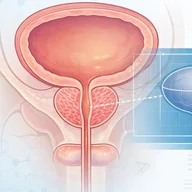
Prostate volume estimation with PSA density calculation using imaging measurements and serum PSA levels.
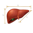
Liver volume estimation using imaging measurements with comparison to expected normal values.

Prostate lesion assessment on MRI using PI-RADS criteria to estimate clinically significant cancer risk.

Splenic volume and index estimation using imaging measurements with reference to normal size ranges.
More about the Ovarian and Testicular Volume Calculators
Clinical Role of the Ovarian Volume Calculator and Testicular Volume Calculator
The Ovarian Volume Calculator and Testicular Volume Calculator provide a standardized approach to gonadal volume estimation across ultrasound, CT, and MRI. In reproductive imaging, gonadal volume serves as a practical surrogate for endocrine function and germ cell activity. In the testis, seminiferous tubules represent the majority of parenchymal volume, which explains the clinical relevance of volumetric assessment in spermatogenesis and hormonal evaluation. Similarly, ovarian volume measurement plays a central role in fertility assessment, follicular monitoring, and ovarian morphology evaluation.
These tools support radiologists and clinicians by applying consistent geometric principles to ovary volume calculation and testis volume calculation. They are designed to complement clinical interpretation and facilitate reproducible measurements in scenarios such as infertility workup, pubertal assessment, and endocrine disorders.
Clinical relevance of ovarian volume and testicular volume assessment
Accurate assessment of ovarian volume and testicular volume is fundamental in longitudinal reproductive care. In scrotal ultrasound testicular measurement, volume is used to evaluate pubertal progression, varicocele-associated hypotrophy, and suspected spermatogenic dysfunction. Clinical estimation methods such as orchidometers tend to overestimate true volume due to inclusion of adjacent structures, whereas imaging-based gonadal volume estimation provides a more controlled measurement environment [4].
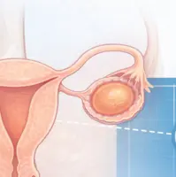
In pelvic ultrasound ovary measurement, ovarian size assessment ultrasound is routinely integrated with follicular assessment. Ovarian morphology and volume contribute to the evaluation of ovarian reserve and differentiation between physiologic multifollicular ovaries and polycystic ovarian morphology. These measurements are commonly interpreted alongside clinical and laboratory parameters.
How to calculate ovarian volume using imaging
Ovarian volume is typically calculated using the standard ellipsoid formula ovary, which applies three orthogonal diameters obtained on imaging:
Volume = Length × Width × Height × 0.523
This approach reflects the geometric approximation of an ellipsoid and is widely used in ovary volume calculation ultrasound workflows. Accurate measurement requires acquisition of the maximum longitudinal, transverse, and anteroposterior dimensions.
How to calculate testicular volume using the ellipsoid formula
Testicular volume is commonly calculated using a modified ellipsoid approach. The ellipsoid formula testis may incorporate different constants depending on methodology:
Volume = Length × Width × Height × 0.71
This formulation, often referred to as the Lambert method, has demonstrated closer agreement with reference standards such as water displacement compared to the traditional 0.523 constant [1]. Awareness of the formula used by ultrasound systems is important, as variations in constants may influence reported testicular size measurement ultrasound values.
| Formula Name | Calculation Method | Typical Use |
|---|---|---|
| Standard Ellipsoid | L × W × H × 0.523 | Ovarian volume estimation |
| Modified Ellipsoid (Lambert) | L × W × H × 0.71 | Testicular volume estimation |
| Hansen Formula | L × W² × 0.52 | Alternative testis volume calculation |
Imaging techniques for gonadal volume estimation
Volumetric estimation can be performed using multiple imaging modalities. Two dimensional ultrasound remains the most commonly used technique, although it relies on geometric assumptions and operator-dependent measurements. Three dimensional ultrasound reduces reconstruction error and enables semi-automated follicular and volume assessment through voxel-based analysis [3].
MRI provides an alternative approach for volumetric estimation, particularly when detailed tissue characterization or reproducibility is required. MRI-based segmentation methods calculate volume directly from voxel counts, avoiding reliance on simplified geometric models.
Ovarian and testicular volume normal ranges and interpretation
Interpretation of ovarian and testicular volume requires consideration of patient age, pubertal stage, and clinical context. In adults, the testicular volume normal range is commonly cited between approximately 14 and 18 mL, although variation exists depending on methodology and population.
Ovarian volume varies across the reproductive lifespan and is influenced by hormonal activity. During reproductive years, ovarian size generally correlates with follicular activity, while reduced volume may be observed with advancing age or diminished ovarian reserve. Serial measurements may provide additional context in longitudinal assessment.
Clinical applications in fertility and endocrine evaluation
Ovarian volume contributes to fertility assessment imaging by supporting evaluation of ovarian reserve and response to stimulation protocols. It is often interpreted alongside antral follicle count and hormonal markers. Related tools such as the O-RADS calculator and pelvic ultrasound interpretation tools may be used in broader adnexal evaluation.
Testicular volume plays a key role in urologic and endocrine assessment, including evaluation of delayed puberty, primary testicular failure, and varicocele-related changes. Integration with hormonal profiles and semen analysis remains essential for comprehensive interpretation.
Additional volumetric tools, such as the Prostate volume and PSA density calculator and other genitourinary radiology calculators, follow similar geometric principles and can support broader clinical workflows.
Limitations of volume estimation in imaging
Several factors influence the accuracy of gonadal volume estimation. Measurement variability is more pronounced in small structures, particularly when volumes are below 4 mL, where interobserver differences may be substantial [2]. Probe positioning, incomplete visualization of maximal diameters, and differences in formula selection can also affect results.
Clinical interpretation should incorporate the full clinical picture, including endocrine markers, developmental stage, and patient history. The Ovarian Volume Calculator and Testicular Volume Calculator are intended to support consistent measurement practices rather than replace clinical judgment.
Frequently Asked Questions (FAQs)
How is ovarian volume calculated on ultrasound?
Ovarian volume is calculated using the ellipsoid formula with three orthogonal measurements. The formula multiplies length, width, and height by 0.523. Accurate measurement depends on identifying the largest dimensions in each plane during pelvic ultrasound ovary measurement.
What is the preferred method for testicular volume calculation?
Testicular volume is commonly calculated using a modified ellipsoid formula with a constant of 0.71. This approach has been shown to align more closely with reference methods compared to the standard ellipsoid constant used in some ultrasound systems.
What is the normal range for testicular volume in adults?
Adult testicular volume typically falls within a range of approximately 14 to 18 mL. Interpretation should consider patient age, clinical context, and measurement technique.
Why is ovarian volume important in fertility assessment?
Ovarian volume is used in conjunction with follicular assessment and hormonal evaluation to estimate ovarian reserve. It provides supportive information in reproductive imaging and assisted reproduction planning.
What are common sources of error in gonadal volume measurement?
Errors may arise from suboptimal probe positioning, incorrect selection of measurement planes, variability between observers, and differences in formula constants used by imaging systems. These factors are particularly relevant in small volume measurements.
References
- Cai D, et al. Validity of measurements of testicular volume obtained by a built-in software of ultrasound systems. J Ultrason. 2020;20(82):e181-e184.
- Schäfer FM, et al. Intra- and Interobserver Variability in Ultrasound Measurement of Testicular Volumes in Pubertal Boys. Children. 2024;11(6):741.
- Vandekerckhove F, et al. The Value of Automated Follicle Volume Measurements in IVF/ICSI. Front Surg. 2014;1:18.
- Hsieh ML, et al. The reliability of ultrasonographic measurements for testicular volume assessment. Asian J Androl. 2009;11(2):261-265.

PGY-5 Radiology and Nuclear Medicine Resident Physician
UT Southwestern Medical Center, USA

Exacto