Bone-RADS Calculator v. 2023
Reference:
Related Calculators
Liver lesion classification for hepatocellular carcinoma risk using CT, MRI, ultrasound, and contrast-enhanced ultrasound.

Thyroid nodule risk stratification using ultrasound features and ACR TI-RADS scoring system.
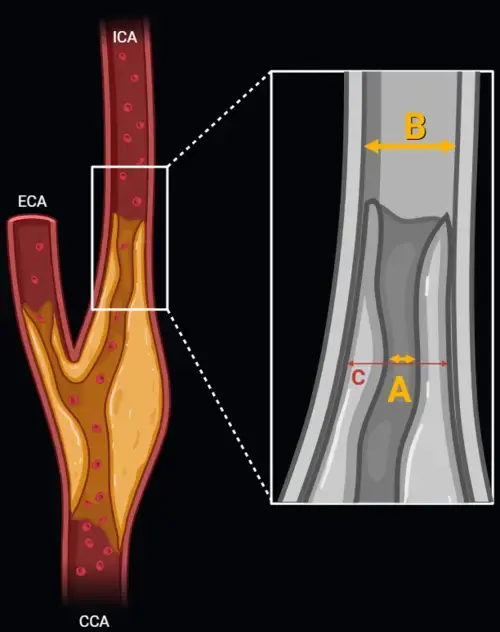
Carotid artery stenosis measurement using NASCET and ECST methods on CT angiography or MR angiography.
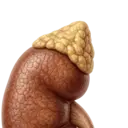
Characterization of adrenal lesions using signal intensity loss on in-phase and opposed-phase MRI sequences.

More about the Bone-RADS Calculator
The Bone Reporting and Data System (Bone-RADS™), introduced by the American College of Radiology (ACR), provides a standardized method for evaluating and managing bone lesions that may be neoplastic. The Bone-RADS calculator helps radiologists assess solitary bone lesions on radiographs, CT, or MRI, offering structured criteria to estimate malignancy risk and guide clinical decisions. This standardized tool improves diagnostic confidence, supports clinical communication, and reduces ambiguity in reporting potentially malignant bone lesions.
Purpose and Use of Bone-RADS™
Bone-RADS™ is designed to be used in the assessment of incidental or suspicious bone lesions discovered during routine imaging. Its goal is to stratify lesions by risk level, allowing radiologists to provide actionable and consistent recommendations. The calculator assigns a Bone-RADS™ category based on key imaging features such as lesion margin, matrix mineralization, periosteal reaction, and associated soft tissue findings. It can be used across various clinical contexts, including orthopedic, oncologic, and emergency imaging scenarios.
Bone-RADS™ Risk Categories and Suggested Actions
The classification system includes four main risk levels:
- Bone-RADS™ 1 – Very Low Risk: Classic benign findings like bone islands (enostoses) or fibrous cortical defects.
Action: No additional imaging or follow-up required. - Bone-RADS™ 2 – Low Risk: Probably benign lesions without conclusive features. Examples include non-aggressive sclerotic or lytic lesions.
Action: Cross-sectional imaging (e.g., MRI) recommended for further characterization. - Bone-RADS™ 3 – Intermediate Risk: Indeterminate lesions, such as well-defined but atypical lucent lesions or those with mild periosteal reaction.
Action: Serial imaging at 6, 6, and 12 months to assess for interval growth or evolution. - Bone-RADS™ 4 – High Risk: Imaging suggests possible malignancy — features like ill-defined margins, aggressive periosteal reactions, or soft tissue mass.
Action: Prompt biopsy and/or referral to orthopedic oncology or musculoskeletal specialists is advised.
Benefits of Using the Bone-RADS Calculator
Integrating the Bone-RADS calculator into radiological practice provides several key benefits:
- Improved Consistency: Ensures uniform lesion classification across institutions and radiologists.
- Streamlined Management: Links imaging appearance directly with clinical recommendations, reducing ambiguity.
- Enhanced Reporting Quality: Facilitates comprehensive and structured radiology reports, aiding referring physicians.
- Educational Resource: Supports learning and professional development for radiologists, residents, and other clinicians involved in musculoskeletal imaging.
Bone-RADS™ in Clinical Context
While Bone-RADS™ is not intended to replace clinical judgment or histopathological analysis, it serves as a valuable adjunct in evaluating bone lesions, particularly those discovered incidentally. The system contributes to better triaging of patients, appropriate use of follow-up imaging, and earlier identification of malignancies requiring intervention.
The Bone-RADS calculator on this page is based on published criteria from the ACR and is designed to be used by trained medical professionals. For additional details, updates, and training materials, visit the official ACR Bone-RADS™ page.

It seems that this calculator incorrectly assigns 4 points for a soft tissue mass, while according to the ACR’s page for Bone-RADS it should assign 2 points only. May you please verify this and fix the bug? Thank you.
Hello Dr. Marek,
Thank you so much for bringing this error to my attention. The bug has been fixed!
I appreciate your feedback.