Radiology Call Resources for Trainees
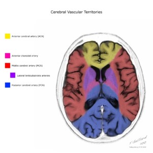
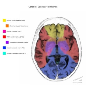
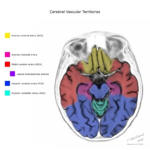
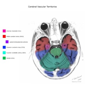
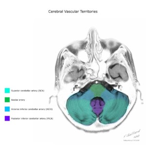
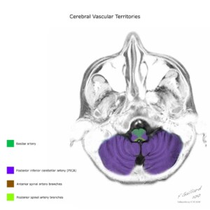
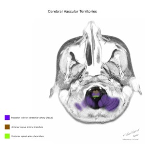
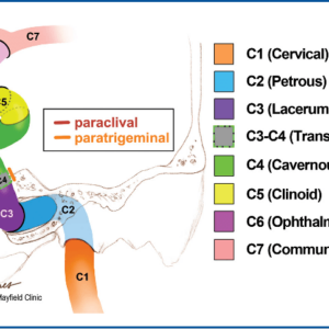
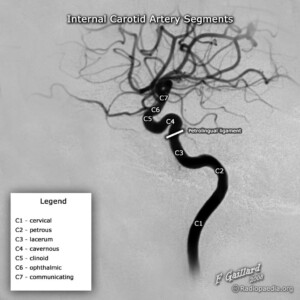
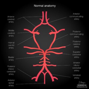
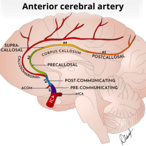
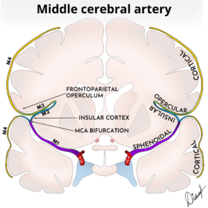
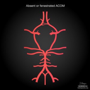
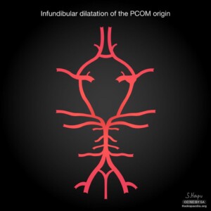
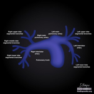
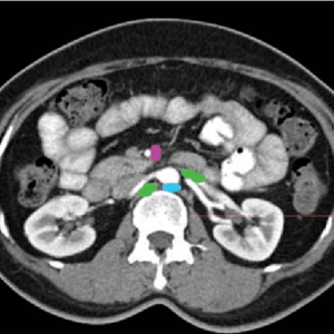
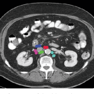
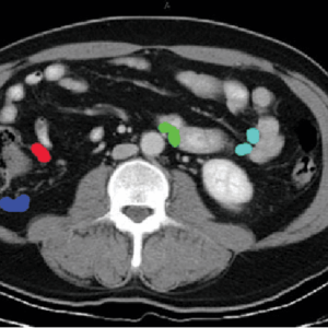
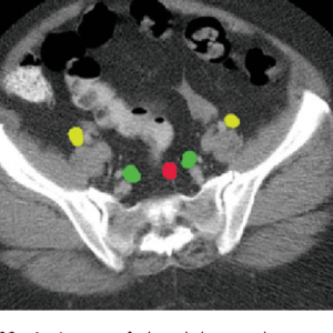
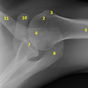
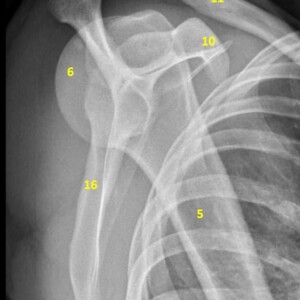
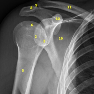
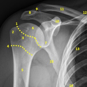
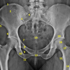
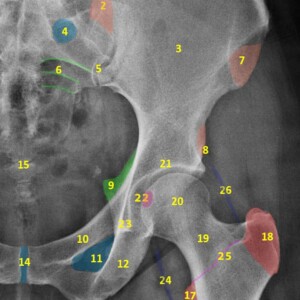
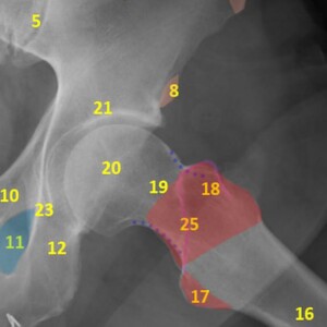
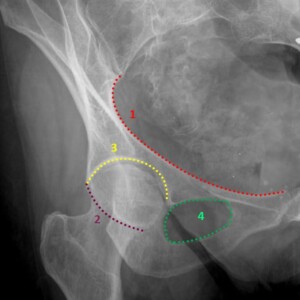
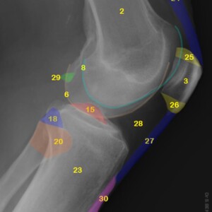
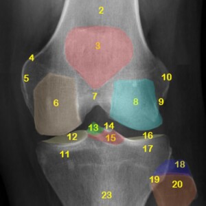
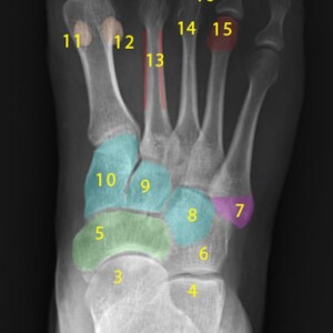
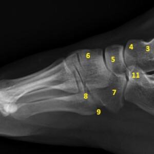
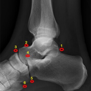
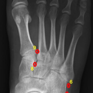
Anatomy
Call Calculators
Trauma injury scoring:
Incidentalomas:
Neuro:
Other:
Diagnostic Parameters
MRI:
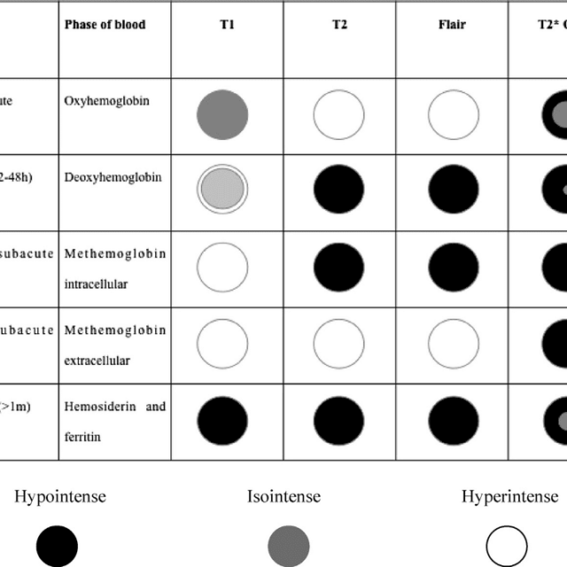
- Staging of brain hemorrhage on MRI
 Appearance of intracerebral hemorrhage on MRI by stageData from Kidwell CS, Chalela JA, Saver JL, et al. Comparison of MRI and CT for detection of acute intracerebral hemorrhage. JAMA 2004;292:1823–30; and Wijman CA, Venkatasubramanian C, Bruins S, et al. Utility of early MRI diagnosis and management of acute spontaneous intracerebral haemorrhage. Cerebrovasc Dis 2010;30:456–63. Image from Domingues, Renan & Rossi, Costanza & Cordonnier, Charlotte. (2015). Diagnostic uation for Nontraumatic Intracerebral Hemorrhage. Neurologic clinics. 33. 315-328. 10.1016/j.ncl.2014.12.001.
Ultrasound:
- Renal Doppler (native & transplant)
Native Renal US Doppler
RI is a nonspecific prognostic marker in vascular diseases that affect the kidney. There is thought to be little correlation between the resistive indices and the quantitative extent of renal dysfunction (measured by serum creatinine values)
- > 0.8: Abnormal. Usually seen with:
- Associated with renal dysfunction and adverse cardiovascular events
- Medical renal disease
- Ureteric obstruction
- Extreme hypotension
- Very young children
- Perinephric fluid collection
- Abdominal compartment syndrome
- 0.7 - 0.8: Indeterminate
- < 0.7: Normal
- < 3.5 Normal
- > 3.5 Abnormal
Normal ranges:
- Aorta: 80-100 cm/sec
- Renal Artery: < 180 cm/sec Normal
- Medullary Artery: 30-40 cm/sec
- Cortical Artery: 20-30 cm/sec
- < 50 msec Normal
- > 50 msec Abnormal
- Normal: Low resistance, Forward diastole flow
- Abnormal: High resistance, Parvus et trades, Monphasic distal to stenosis
Transplant Renal US Doppler
RI is a nonspecific prognostic marker in vascular diseases that affect the kidney. There is thought to be little correlation between the resistive indices and the quantitative extent of renal dysfunction (measured by serum creatinine values)
- > 0.8: Abnormal (More specific for disease than low RI). Associated with increased risk of graft loss and death. Usually seen with:
- Drug toxicity
- Acute/Chronic transplant rejection
- Acute tubular necrosis (ATN)
- Renal vein thrombosis (a specific waveform)
- Graft infection
- Compressive perinephric fluid collections
- Obstructive hydronephrosis
- 0.7 - 0.8: Indeterminate
- 0.5 - 0.7: Normal
- < 0.5: Sometimes abnormal (Less specific for disease than high RI)
RAS usually happens more than 3 months post-transplant and presents with HTN. Risk factors are live donors and children. The combination of both direct and indirect Doppler measurements gives an overall accuracy of 95%.
Diagnostic criteria:
- Peak Systolic Velocity (PSV)
- > 200 cm/sec
- No significant stenosis if RIR < 2.0 AND no tradus-parvus (If elevated PSV is the only finding, it's not fully reliable
- Tortuous Vessels
- Transplant kidney has more tortuous vessels than native renal arteries
- Flow normally accelerates around curves or kinks
- Suspect increased velocity is due to tortuosity when:
- PSV > 200 cm/s
- RIR ≥2.0
- Curvy vessel/small angle
- Absent tardus parvus
- Renal Iliac Ratio (RIR)
- > 2.0
- Ratio of the highest PSV of the main renal artery (proximal, mid, or distal) to the PSV of the iliac artery proximal to the anastomosis
- Marked Distal Turbulence
- Spectral broadening
- Tardus-parvus Segmental Branch Waveform
- Indirect sign of a significant (> 80%) proximal arterial stenosis
- Prolonged acceleration time > 0.07 sec
- Decreased RI < 0.56
- Loss of early systolic peak
Rare (< 1%) condition that happens immediately post-op/intra-op. Risk factors are Living donor, Complex arterial anastomoses, Pediatric (small vessel size), Severe rejection, and ATN.
Doppler US Findings
- Absent arterial AND venous flow
- Pulsed wave Doppler is MORE sensitive than either color or power Doppler
Rare (< 5%) condition that usually happens less than 1 week post-op, and presents with decreased urine output, swelling, and tenderness over the graft. Risk factors are left lower quadrant allografts, surgical difficulty with the venous anastomosis, episodes of hypovolemia, venous compression by a peritransplant collection, and slow flow secondary to rejection.
Doppler US Findings
- Diagnostic Criteria
- Reversed diastolic flow in the main renal artery and/or intrarenal arteries
- Reduced or no flow in the main renal vein
- Additional Findings
- Large size
- Hypoechoic with loss of corticomedullary differentiation
- Echogenic material may be in the renal vein
- If thrombosis is partial, high RI may be seen
- Hematoma
- Seroma
- Lymphoceles
- Abscess
- Urinoma
Happens in 2% of cases, usually (> 90%) within the distal third of the ureter and due to relatively poor blood supply. Strictures are usually observed at the ureterovesical junction.
Risk factors are Ischemia or rejection, Surgical technique, Kinking, peritransplant fluid collections
Presents with rising level of serum creatinine
Other causes of mild dilatation of the collecting system:
- Chronic rejection
- Normal finding in the early transplant kidney, due to tonicity loss secondary to denervation and increased flow through the single functioning kidney
- A distended bladder alone can be the underlying cause
- Perinephric fluid collections that may cause ureteral compression
References: MobiBrain, doi:10.5402/2013/480862, and doi:10.1016/j.jus.2009.09.006 - > 0.8: Abnormal. Usually seen with:
- Viability of first trimester pregnancy
Findings diagnostic of pregnancy failure
Crown-rump length (CRL) of ≥7 mm and no heartbeat on a transvaginal scan
Mean sac diameter (MSD) of ≥25 mm and no embryo on a transvaginal scan
Absence of embryo with heartbeat ≥2 weeks after a scan that showed a gestational sac without a yolk sac
Absence of embryo with heartbeat ≥11 days after a scan that showed a gestational sac with a yolk sac
Sac with no embryo and an MSD <12 mm on initial scan that fails to double in size on a scan ≥14 days later
Sac with no embryo and an MSD ≥12 mm on initial scan with no embryo heart activity on a scan ≥7 days later
Embryo (irrespective of crown-rump length) without cardiac activity on initial scan and on repeat scan ≥7 days later
Cessation of a previously documented cardiac activity of embryo (irrespective of crown-rump length)
Findings suspicious but not diagnostic of pregnancy failure
Crown-rump length (CRL) of <7 mm and no heartbeat
Mean sac diameter (MSD) of 16-24 mm and no embryo
Absence of embryo with heartbeat 7-13 days after a scan that showed a gestational sac without a yolk sac
Absence of embryo with heartbeat 7-10 days after a scan that showed a gestational sac with a yolk sac
Absence of embryo ≥6 weeks after last menstrual period
Absence of embryo when amnion seen adjacent to yolk sac (empty amnion sign)
Embryo present with amnion visible around it but no heartbeat (expanded amnion sign)
Yolk sac that is separated from an embryo when CRL is ≤ 5 mm (yolk stalk sign)
Small gestational sac in relation to the size of the embryo (<5 mm difference between mean sac diameter and crown-rump length)
Enlarged yolk sac (>7 mm)
Reference: Jones J, Yu Y, Yap J, et al. Failed early pregnancy. https://doi.org/10.53347/rID-9040 - Arteriovenous fistula/graft Doppler (coming soon)
- Carotid Doppler (coming soon)
- Mesenteric Doppler (coming soon)
- Limb Doppler (upper and lower extremities + ABI/WBI) (coming soon)
- Liver Doppler (native, transplant, and TIPS) (coming soon)
General:
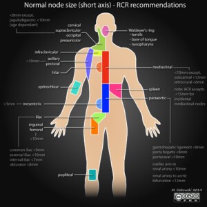
- Normal sizes in adults (coming soon)
- Normal sizes in children (coming soon)

