Splenic Index Calculator & Spleen Volume Assessment (US, CT, MRI)
Measurement guidelines for CT & Ultrasound
Related Calculators
Liver volume estimation using imaging measurements with comparison to expected normal values.

Prostate volume estimation with PSA density calculation using imaging measurements and serum PSA levels.

Thyroid nodule risk stratification using ultrasound features and ACR TI-RADS scoring system.
Gonadal volume estimation using imaging measurements for evaluation of size, development, and pathology.
More about the Splenic Index and Volume Calculator
Introduction: Quantitative Spleen Assessment in Modern Imaging
The Splenic Index Calculator provides a structured method for quantitative splenic size assessment using cross sectional imaging or ultrasound measurements. In routine radiology practice, objective organ size assessment has largely replaced reliance on physical examination alone, since clinical palpation detects splenic enlargement only after substantial volumetric increase [1].
By applying standardized measurement techniques, the Splenic Index Calculator and the complementary Spleen Volume Calculator support reproducible documentation of spleen dimensions radiology reports. These tools are intended to assist clinical decision making and longitudinal comparison, particularly in splenomegaly assessment imaging workflows.
Clinical Significance of Splenomegaly on Imaging
Splenomegaly reflects a broad differential diagnosis across infectious, hematologic, inflammatory, congestive, and infiltrative processes. Accurate quantification improves diagnostic clarity and follow up assessment.
Infectious and inflammatory causes include viral infections such as infectious mononucleosis, as well as malaria, leishmaniasis, tuberculosis, brucellosis, and bacterial endocarditis [1].
Hematologic disorders commonly associated with splenic enlargement include lymphoma, leukemia, hemolytic anemia, myelofibrosis, and polycythemia vera [1].
Congestive and infiltrative etiologies include portal hypertension secondary to cirrhosis, amyloidosis, sarcoidosis, and storage disorders [1].
Hypersplenism represents a clinical syndrome of cytopenias related to splenic sequestration and enlargement. Imaging based organ size assessment should always be interpreted in conjunction with laboratory findings and hepatic morphology.
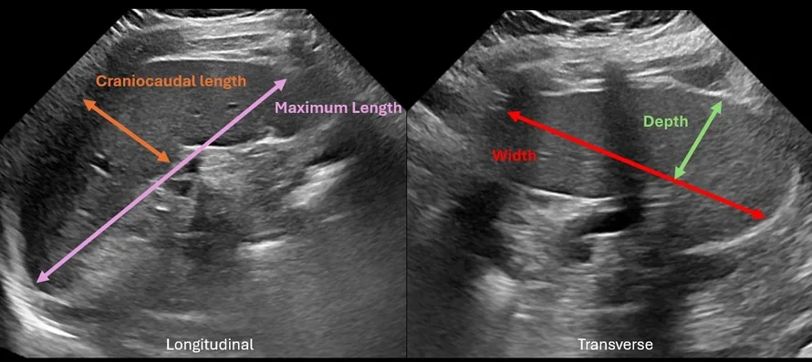
How to Calculate the Splenic Index
The splenic index formula is defined as:
Splenic Index = Length × Width × Thickness
Measurements are obtained as follows:
- Length: Maximum longitudinal dimension, often best captured on oblique coronal CT reformations.
- Width: Maximum dimension at the hilum on axial imaging.
- Thickness: Maximum anteroposterior dimension perpendicular to width.
The calculated value approximates splenic volume in cubic centimeters. Published reference ranges commonly cite values between 120 and 480 cm³ in adults [2]. Interpretation should account for patient habitus and clinical context.
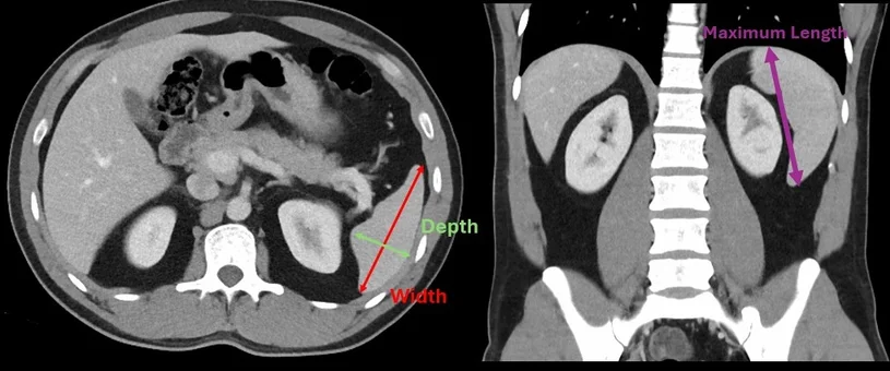
Spleen Volume Calculator and CT Splenic Volumetry
The Spleen Volume Calculator applies the prolate ellipsoid formula to better approximate three dimensional morphology during spleen volume measurement CT evaluation:
Volume = 0.524 × (Length × Width × Thickness)
An alternative approach averages maximum longitudinal length and strict craniocaudal length to better reflect curved morphology:
Volume = 0.524 × Width × Thickness × ((Maximum Length + Craniocaudal Length) / 2) [2]
Reported normal adult splenic volumes typically range from 26 to 250 cm³, though population studies demonstrate wide variability [2]. Volumetric spleen analysis may be particularly helpful for borderline enlargement or therapy monitoring.
| Metric | Typical Adult Range | Common Threshold Suggesting Splenomegaly |
|---|---|---|
| Craniocaudal Length | Up to 12 cm | ≥ 13 cm |
| Splenic Index | 120 to 480 cm³ | > 480 cm³ |
| Splenic Volume | 26 to 250 cm³ | > 250 cm³ |
Population Variability and Anthropometric Considerations
Normal spleen size ultrasound and CT measurements correlate with age, sex, height, and body mass index. Quantitative CT studies demonstrate significant variability in healthy adults, with mean volumes near 212 cm³ and broad standard deviations [1]. Interpretation should therefore integrate anthropometric context rather than relying on a single fixed cutoff.
Splenic Index Versus CT Volumetry
Linear measurements are efficient and widely reproducible but may underestimate transverse or irregular enlargement.
Splenic index calculations incorporate three dimensional measurement and improve correlation with overall splenic mass compared with length alone.
CT splenic volumetry using the ellipsoid constant provides a closer geometric approximation of organ morphology and may support longitudinal assessment in hematologic disease.
Imaging Evaluation of Splenomegaly
Technical precision is essential for accurate splenomegaly assessment imaging:
- Identify the notched anterior border to distinguish spleen from adjacent structures [3].
- Exclude accessory spleens from primary volumetric calculations.
- Use multiplanar reformations to ensure maximal craniocaudal length capture.
- Avoid measurement distortion from rib overlap or oblique slice orientation.
For comprehensive abdominal assessment, clinicians may also reference related abdominal measurement calculators, portal hypertension imaging tools, liver volume calculator resources, and other radiology organ volumetry tools.
Role of Quantitative Tools in Clinical Practice
The Splenic Index Calculator and Spleen Volume Calculator function as structured decision support resources. Their outputs depend on correct application of established measurement techniques and careful image review. Final interpretation remains grounded in comprehensive radiologic evaluation and clinical correlation.
Frequently Asked Questions (FAQs)
What is the normal splenic index in adults?
Commonly cited adult reference ranges fall between 120 and 480 cm³, though values vary with body habitus and demographic factors [2].
How does the splenic index compare with CT volumetry?
The splenic index provides a three dimensional linear approximation. CT volumetry using the ellipsoid constant more closely models organ geometry and may offer improved consistency for follow up comparison [2].
When is volumetric spleen analysis preferred?
Volumetric assessment may be helpful in borderline enlargement, research settings, or longitudinal monitoring of hematologic disorders.
Does BMI affect spleen size interpretation?
Yes. Published CT studies demonstrate correlations between splenic volume and anthropometric variables including BMI and age [1].
What are common pitfalls in spleen volume measurement CT?
Frequent issues include inaccurate plane selection, failure to identify accessory spleens, and misidentification of adjacent structures due to anatomic crowding [3].
References
- Mukhtar MA. Determination of normal splenic dimensions on computed tomography images in Sudanese population. Sudan University of Science and Technology; 2016. Available at: https://repository.sustech.edu/handle/123456789/13916
- Loftus WK, Chow LT, Metreweli C. Sonographic measurement of splenic length: correlation with splenic volume. AJR Am J Roentgenol. 1999;172(2):455-458. Available at: https://www.ajronline.org/doi/full/10.2214/ajr.172.2.9930794
- Standring S, ed. Gray’s Anatomy: The Anatomical Basis of Clinical Practice. 39th ed. Elsevier; 2005. Available at: https://www.elsevier.com/books/grays-anatomy/standring/978-0-443-06684-9

PGY-5 Radiology and Nuclear Medicine Resident Physician
UT Southwestern Medical Center, USA
