
Duke 4D CT Parathyroid Calculator for Adenoma & Hyperplasia
Related Calculators
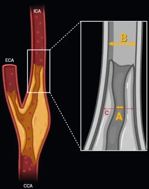
Carotid artery stenosis measurement using NASCET and ECST methods on CT angiography or MR angiography.
Standardized spinal trauma classification based on injury morphology, neurological status, and additional modifiers.
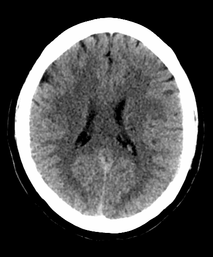
Early ischemic stroke assessment using CT scoring of middle cerebral artery territory involvement.

MRI-based grading of cavernous sinus invasion using the Knosp classification for pituitary tumors
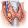
More about the Duke 4D CT Parathyroid Adenoma and Hyperplasia Enhancement Pattern Calculator
The Duke 4D CT parathyroid calculator is a structured reference for evaluating candidate parathyroid lesions on multiphase CT. The framework combines enhancement relative to the thyroid gland with selected morphologic findings to categorize lesions using the Duke 4D-CT scoring system.[1][2] It is intended to support systematic image interpretation rather than replace review of the complete examination, clinical history, biochemical findings, or operative correlation.
In patients with primary hyperparathyroidism being evaluated for surgery, 4D CT parathyroid adenoma localization uses both anatomy and changes in contrast enhancement over time. This can be particularly useful when evaluating ectopic lesions, small candidate glands, multigland disease, or findings that are difficult to characterize with other localization studies.[3][4]
How parathyroid 4D CT evaluates enhancement over time
The "fourth dimension" in parathyroid 4D CT is time. Multiphase imaging allows the radiologist to compare a candidate lesion with the thyroid gland before and after intravenous contrast administration. Protocols vary among institutions, but commonly include noncontrast, arterial, and delayed contrast-enhanced phases.[1][3]
On noncontrast imaging, abnormal parathyroid tissue is often lower in attenuation than the iodine-rich thyroid gland. Following contrast administration, many parathyroid lesions demonstrate rapid enhancement, washout, or another enhancement pattern that differs from the adjacent thyroid. The relative relationship to the thyroid is central to the Duke framework and is generally more useful than relying on a single absolute Hounsfield unit threshold.[1][2]
Duke three-pattern framework for parathyroid adenoma and hyperplasia
The three enhancement patterns were described by Bahl, Sepahdari, Sosa, and Hoang in a study of pathologically proven parathyroid adenomas and hyperplastic glands.[1] All three patterns begin with a candidate lesion that is lower in attenuation than the thyroid gland on the noncontrast phase. The types are then distinguished by the lesion's relationship to the thyroid on arterial and delayed imaging.
| Enhancement type | Noncontrast phase | Arterial phase | Delayed phase | Interpretive concept |
|---|---|---|---|---|
| Type A | Lower attenuation than thyroid | Higher attenuation than thyroid | May demonstrate subsequent washout | Arterial hyperenhancement relative to the thyroid is the defining feature of the original Type A pattern |
| Type B | Lower attenuation than thyroid | Not higher attenuation than thyroid | Lower attenuation than thyroid | Delayed hypoattenuation relative to the thyroid identifies the lesion despite the absence of Type A arterial hyperenhancement |
| Type C | Lower attenuation than thyroid | Not higher attenuation than thyroid | Not lower attenuation than thyroid | The postcontrast relationship to the thyroid is less distinctive, increasing reliance on morphology and anatomic location |
Type A is often associated with the familiar concept of rapid arterial enhancement, while Type B illustrates why a candidate parathyroid lesion does not need to become brighter than the thyroid during the arterial phase. Type C lesions are especially important because their postcontrast attenuation may overlap more substantially with the thyroid or other cervical structures.[1]
This distinction is useful when interpreting a parathyroid adenoma enhancement pattern. A lesion should not be excluded solely because it lacks dramatic arterial hyperenhancement. Comparison across all available phases, together with location and morphology, is more informative than expecting every abnormal gland to demonstrate a single classic "fast-in, fast-out" appearance.
How the Duke parathyroid scoring system combines enhancement and morphology
A later description of the Duke 4D-CT scoring system combines the enhancement type with three secondary imaging findings: lesion size of at least 1 cm, a polar vessel, and cystic change.[2] These findings are used to place a candidate lesion into one of three confidence categories.
| Enhancement pattern | Secondary finding | Published Duke category |
|---|---|---|
| Type A or Type B | At least one secondary finding | Consistent with |
| Type A or Type B | No secondary finding | Highly suspicious |
| Type C | At least one secondary finding | Highly suspicious |
| Type C | No secondary finding | Possible |
The terms "consistent with," "highly suspicious," and "possible" are category labels used in the published Duke framework.[2] They should not be interpreted as histopathologic diagnoses or as guarantees that a candidate lesion represents hyperfunctioning parathyroid tissue.
Secondary imaging features used in the Duke 4D CT scoring system
Size of at least 1 cm: Lesion size is incorporated as one of the secondary findings. Size should be considered together with enhancement and morphology rather than used as an isolated diagnostic threshold.
Polar vessel sign: A visible feeding or draining vessel entering one pole of a candidate lesion can support a parathyroid origin. This finding is useful as an adjunct but should be interpreted in the context of the lesion's location and enhancement characteristics.[2][3]
Cystic change: Internal cystic change is another secondary feature incorporated into the Duke scoring framework. Cystic lesions may demonstrate less typical enhancement because only the residual solid tissue participates in the expected contrast kinetics.[2]
4D CT parathyroid hyperplasia and multigland disease
The enhancement-pattern framework is relevant to both adenomas and hyperplastic parathyroid glands. The original Duke study included lesions representing both parathyroid adenoma and hyperplasia.[1] This is important because 4D CT parathyroid hyperplasia and multigland disease may present differently from a single dominant adenoma.
In multigland parathyroid disease, individual abnormal glands may be smaller and less conspicuous. Identification of one convincing lesion therefore should not automatically end the search. The remainder of the expected parathyroid locations and common ectopic pathways should still be reviewed for additional candidate lesions.[4][5]
Parathyroid washout on 4D CT
The term "washout" describes decreasing lesion enhancement after an earlier contrast-enhanced phase. It is commonly associated with parathyroid adenomas, but the Duke framework demonstrates that abnormal parathyroid glands do not all follow the same enhancement curve.[1] For this reason, parathyroid washout on 4D CT should be interpreted relative to the thyroid gland and in conjunction with the complete multiphase appearance.
Absolute attenuation values can also vary with scanner technique, contrast administration, cardiac output, lesion composition, and acquisition timing. Relative enhancement patterns can therefore provide a useful framework for interpretation without requiring a universal Hounsfield unit cutoff.
Common mimics of a parathyroid adenoma on 4D CT
Thyroid nodules and cervical lymph nodes are among the most important mimics of abnormal parathyroid tissue. Enhancement alone is not always sufficient to distinguish these structures. Evaluation should incorporate noncontrast attenuation, enhancement across phases, shape, location, relationship to the thyroid, and vascular features.[2][3]
Intrathyroidal or immediately juxtathyroidal lesions can be particularly challenging because thyroid nodules may closely resemble parathyroid tissue. Conversely, lymph nodes may demonstrate enhancement characteristics that overlap with less typical parathyroid lesions. Type C candidate lesions deserve particular attention to these additional features because their postcontrast relationship to the thyroid is less distinctive.
Other pitfalls when interpreting parathyroid 4D CT
Several factors can alter the expected appearance of a candidate lesion. Thyroid heterogeneity or thyroiditis can reduce the usefulness of the thyroid as an internal attenuation reference. Cystic change, hemorrhage, small lesion size, contrast timing, streak artifact, and unusual vascular supply may also produce atypical enhancement.
Ectopic glands may occur outside the expected posterior thyroid locations, including the tracheoesophageal groove, retropharyngeal region, carotid space, thymic pathway, and mediastinum. A systematic search pattern remains important even when the enhancement characteristics of the initial candidate lesion appear convincing.
Practical use in radiology reporting
When a candidate parathyroid lesion is identified, the report can describe its side, superior or inferior location when reasonably determined, dimensions, relationship to the thyroid and nearby landmarks, enhancement type, relevant secondary features, and whether additional candidate lesions are present. In ectopic cases, precise description of the relationship to the trachea, esophagus, carotid vessels, thyroid gland, or mediastinum may be more useful than forcing the lesion into a conventional quadrant.
The Duke 4D CT parathyroid scoring system provides structured terminology for organizing these findings. The final interpretation should still incorporate the entire imaging examination and the clinical and biochemical context. A calculator can assist with assigning the published category from entered imaging features, but it does not determine pathology or establish the diagnosis of primary hyperparathyroidism.
Limitations and safety considerations
4D CT uses ionizing radiation, and radiation exposure varies according to scanner parameters, coverage, patient size, and the number of acquired phases. Because most 4D CT protocols also use intravenous iodinated contrast, patient-specific contrast considerations should be reviewed according to local clinical protocols.
Another limitation is that enhancement patterns are not unique to parathyroid tissue. Small lesions, multigland disease, cystic lesions, intrathyroidal lesions, and Type C enhancement may be more difficult to distinguish from surrounding structures. The scoring framework is therefore best used as an organizational aid for imaging findings rather than as a standalone diagnostic test.
Frequently Asked Questions (FAQs)
What is the Duke 4D CT scoring system for parathyroid lesions?
The Duke 4D-CT scoring system combines a candidate lesion's Type A, B, or C enhancement pattern with secondary features including size of at least 1 cm, a polar vessel, and cystic change. These findings are used to assign the published categories "consistent with," "highly suspicious," or "possible."[2]
What are the three Duke enhancement patterns on parathyroid 4D CT?
All three patterns begin with a lesion that is lower in attenuation than the thyroid on noncontrast imaging. Type A demonstrates arterial attenuation higher than the thyroid. Type B is not higher than the thyroid in the arterial phase but becomes lower than the thyroid on delayed imaging. Type C meets neither the Type A nor Type B postcontrast criteria.[1]
Does a parathyroid adenoma always show arterial hyperenhancement and delayed washout?
No. Although arterial hyperenhancement and subsequent washout are familiar features of parathyroid adenomas, the Duke enhancement framework was developed specifically because abnormal parathyroid glands demonstrate more than one enhancement pattern.[1]
Why is the noncontrast phase useful in parathyroid 4D CT?
The normal thyroid is relatively high in attenuation on noncontrast CT because of its intrinsic iodine content. Candidate parathyroid lesions in the Duke framework are lower in attenuation than the thyroid on this phase, providing an important baseline before contrast enhancement is assessed.[1][3]
What imaging features are used in addition to enhancement pattern?
The Duke scoring framework incorporates three secondary findings: lesion size of at least 1 cm, a polar vessel, and cystic change. Location, shape, relationship to the thyroid, and the presence of additional candidate lesions should also be considered during full image interpretation.[2]
Can the Duke 4D CT framework be applied to parathyroid hyperplasia?
The original enhancement-pattern study included both parathyroid adenomas and hyperplastic glands. In suspected multigland disease, the reader should continue evaluating for additional abnormal glands rather than relying only on the most conspicuous lesion.[1][4]
References
- Bahl M, Sepahdari AR, Sosa JA, Hoang JK. Parathyroid adenomas and hyperplasia on four-dimensional CT scans: three patterns of enhancement relative to the thyroid gland justify a three-phase protocol. Radiology. 2015;277(2):454-462. doi:10.1148/radiol.2015142393. PMID: 26024308.
- Malinzak MD, Sosa JA, Hoang J. 4D-CT for detection of parathyroid adenomas and hyperplasia: state of the art imaging. Current Radiology Reports. 2017;5(2):8. doi:10.1007/s40134-017-0198-8.
- Hoang JK, Sung WK, Bahl M, Phillips CD. How to perform parathyroid 4D CT: tips and traps for technique and interpretation. Radiology. 2014;270(1):15-24. doi:10.1148/radiol.13122661. PMID: 24354373.
- Yeh R, Tay YKD, Tabacco G, et al. Diagnostic performance of 4D CT and sestamibi SPECT/CT in localizing parathyroid adenomas in primary hyperparathyroidism. Radiology. 2019;291(2):469-476. doi:10.1148/radiol.2019182122. PMID: 30835187.
- Rodgers SE, Hunter GJ, Hamberg LM, et al. Improved preoperative planning for directed parathyroidectomy with 4-dimensional computed tomography. Surgery. 2006;140(6):932-940. doi:10.1016/j.surg.2006.07.028. PMID: 17188140.

Radiologist and Nuclear Medicine Specialist
Neuroradiology fellow at Johns Hopkins University
UT Southwestern Medical Center alumnus