
Bern Score Calculator for Spontaneous Intracranial Hypotension
Related Calculators
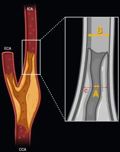
Carotid artery stenosis measurement using NASCET and ECST methods on CT angiography or MR angiography.
Standardized spinal trauma classification based on injury morphology, neurological status, and additional modifiers.
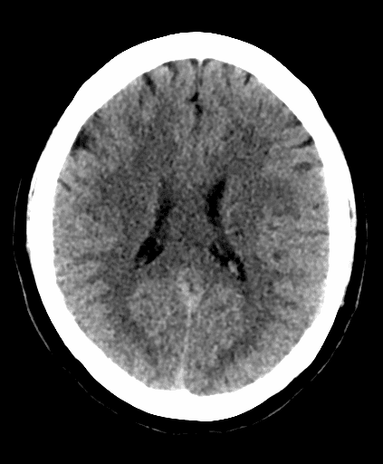
Early ischemic stroke assessment using CT scoring of middle cerebral artery territory involvement.

Estimate intracerebral hemorrhage volume on CT using the ABC/2 method and ellipsoid formula
More about the Bern Score Calculator for Spontaneous Intracranial Hypotension
The Bern Score for spontaneous intracranial hypotension is a 9-point brain MRI scoring system that combines six imaging findings associated with spinal cerebrospinal fluid loss. Developed by Dobrocky and colleagues, the score stratifies patients with suspected spontaneous intracranial hypotension (SIH) into low, intermediate, or high probability groups for identifying a spinal CSF leak on subsequent spinal imaging.[1] It provides a structured approach to SIH brain MRI interpretation but does not independently establish the diagnosis of SIH or identify the site or mechanism of a leak.
The score incorporates imaging manifestations of reduced intracranial CSF volume, including venous engorgement, pachymeningeal enhancement, subdural collections, and downward displacement or crowding of intracranial structures. These findings are interpreted together because no single brain MRI feature is present in every patient with SIH.[1][7]
What the Bern Score measures on brain MRI
The Bern Score SIH framework includes three major criteria worth 2 points each and three minor criteria worth 1 point each. The maximum possible score is 9.[1] Contrast-enhanced brain MRI is required to assess pachymeningeal enhancement and is typically interpreted together with sagittal and multiplanar imaging for evaluation of the basal cisterns and venous structures.
| Criterion | MRI definition | Points |
|---|---|---|
| Pachymeningeal enhancement | Diffuse smooth pachymeningeal enhancement on postcontrast T1-weighted imaging | 2 |
| Venous sinus engorgement | Distension of the dural venous sinuses, including a convex inferior contour of the dominant transverse sinus associated with the venous distension sign | 2 |
| Suprasellar cistern 4.0 mm or less | Reduced suprasellar cistern dimension reflecting downward displacement and crowding of the suprasellar structures | 2 |
| Subdural fluid collection | Presence of a subdural hygroma or hematoma | 1 |
| Prepontine cistern 5.0 mm or less | Reduced distance between the ventral pons and clivus | 1 |
| Mamillopontine distance 6.5 mm or less | Reduced distance between the mamillary bodies and the superior surface of the pons | 1 |
The cisternal measurements are intended to quantify elements of the brain sagging pattern that can accompany spinal CSF loss. Measurement technique and plane selection should be consistent, particularly for values close to a threshold. Other findings associated with spontaneous intracranial hypotension MRI, such as pituitary enlargement or additional manifestations of brain sagging, may support the imaging impression but are not independently scored in the original Bern system.[1]
How the Bern Score is interpreted
The total score is divided into three probability categories. Importantly, these categories describe the published probability of identifying a spinal CSF leak on further imaging rather than the certainty of an SIH diagnosis.[1]
| Total score | Published category | Interpretation |
|---|---|---|
| 0 to 2 | Low probability | Lower probability of identifying a spinal CSF leak on subsequent spinal imaging |
| 3 to 4 | Intermediate probability | Intermediate probability of identifying a spinal CSF leak |
| 5 to 9 | High probability | Higher probability of identifying a spinal CSF leak on subsequent imaging |
A high Bern Score can therefore increase suspicion for an underlying spinal leak and may help inform decisions about further investigation. It should not be interpreted as confirmation of a leak, and the score itself does not determine whether CT myelography, digital subtraction myelography, or another technique should be performed.[1][6]
Why these MRI findings occur in spontaneous intracranial hypotension
Many brain MRI findings of SIH can be understood through changes in intracranial volume following spinal CSF loss. Reduction in CSF volume is accompanied by compensatory expansion of other intracranial compartments, particularly the venous compartment. This contributes to venous sinus engorgement and pachymeningeal enhancement.
Loss of normal CSF buoyancy may also permit downward displacement of intracranial structures. This brain sagging pattern contributes to narrowing of the suprasellar and prepontine cisterns and reduction of the mamillopontine distance. Subdural hygromas or hematomas may occur as another manifestation of altered intracranial mechanics.
Bern Score versus Brain SIH Score
The terminology Brain SIH Score, often abbreviated bSIH Score, appears in later work from the Bern group evaluating brain MRI after treatment. That study used the same six MRI findings, the same weighting, and the same 0 to 9 scale as the original scoring system.[2]
The distinction is primarily one of application. The original scoring system was developed to stratify the likelihood of finding a spinal CSF leak in patients investigated for SIH.[1] The later bSIH publication evaluated the score as an imaging marker before and after surgical closure of a proven spinal dural leak.[2] For a calculator implementing the six original criteria, the underlying point calculation remains the same.
Bern Score and spinal CSF leak imaging
The Bern Score does not localize a leak. Its principal role is to summarize intracranial MRI findings that may alter the pretest probability of detecting spinal CSF loss. Depending on the overall clinical scenario and spine MRI findings, subsequent evaluation may include CT myelography, dynamic CT myelography, or digital subtraction myelography.[6]
Later studies have shown that higher Bern Scores are associated with greater diagnostic yield from specialized myelographic examinations.[3][4] The appropriate myelographic technique also depends on whether spine MRI demonstrates a spinal longitudinal extradural CSF collection and on the suspected mechanism of leakage.
Bern Score and CSF venous fistula
A CSF venous fistula is a spinal connection through which CSF passes directly into the venous system without necessarily producing an extradural CSF collection. This mechanism can make localization particularly challenging and has increased the use of lateral decubitus dynamic imaging techniques.
In a retrospective study of 48 selected patients with suspected SIH and no extradural fluid collection on spine MRI, the Bern Score was strongly associated with detection of a CSF venous fistula using lateral decubitus dynamic CT myelography with bolus tracking.[4] These results are specific to that study population and imaging protocol and should not be interpreted as universal performance characteristics of the Bern Score or this calculator.
A low Bern Score does not exclude SIH
A common interpretation pitfall is treating a low score or a relatively normal brain MRI as evidence against SIH. Patients with proven spinal CSF leaks can have few or no intracranial MRI abnormalities. Normal brain MRI findings have also been reported in systematic reviews of patients with SIH.[7]
This distinction is especially important when clinical suspicion remains high. The decision to pursue further spinal imaging should therefore incorporate symptoms, previous imaging, spine MRI findings, treatment history, and specialist assessment rather than relying exclusively on the numerical score.[6]
The Bern Score does not measure clinical severity
The number of MRI abnormalities does not necessarily parallel the severity of a patient's headache or functional impairment. Houk and colleagues found only a low correlation between pretreatment Bern Scores and headache severity measured with the Headache Impact Test-6.[5]
For this reason, the score should not be used as a surrogate for symptom burden, disability, treatment eligibility, or the severity of the patient's experience. Its purpose is imaging based probability stratification.
Important interpretation pitfalls
Individual Bern Score findings are not specific to SIH. Pachymeningeal enhancement has a differential diagnosis that includes infectious, inflammatory, neoplastic, and postprocedural processes. Subdural collections also have numerous potential causes. Venous sinus contour and basal cistern dimensions should be interpreted within the overall anatomy and clinical context.
Timing also matters. SIH imaging findings may evolve during the disease course or after treatment. A prior epidural blood patch, spontaneous change in the leak, or definitive treatment can alter the appearance of the brain MRI. Comparison with previous studies can therefore be useful when available.
Practical application in radiology reporting
When the Bern Score is used in clinical practice, a radiology report can document the individual positive MRI findings, relevant cisternal measurements, and resulting probability category. The score is most useful when accompanied by a conventional imaging impression rather than presented as an isolated number.
A Bern Score calculator provides a structured method for applying the published point system. The calculated category should be interpreted together with the complete brain MRI, spine imaging, clinical presentation, and subsequent diagnostic evaluation. The calculator does not independently diagnose spontaneous intracranial hypotension or determine the location of a spinal CSF leak.
Frequently Asked Questions (FAQs)
What is the Bern Score for spontaneous intracranial hypotension?
The Bern Score is a 9-point brain MRI scoring system that combines six imaging findings to classify patients into low, intermediate, or high probability groups for identifying a spinal CSF leak on subsequent imaging.[1]
Which MRI findings are major Bern Score criteria?
The three major criteria are pachymeningeal enhancement, venous sinus engorgement, and a suprasellar cistern measurement of 4.0 mm or less. Each major criterion contributes 2 points.[1]
Which findings are minor Bern Score criteria?
The minor criteria are a subdural fluid collection, a prepontine cistern measurement of 5.0 mm or less, and a mamillopontine distance of 6.5 mm or less. Each contributes 1 point.[1]
What does a high Bern Score mean?
A score of 5 to 9 is categorized as high probability and is associated with a greater likelihood of identifying a spinal CSF leak on subsequent spinal imaging. It does not by itself confirm SIH or a specific leak mechanism.[1]
Can spontaneous intracranial hypotension occur with a low Bern Score?
Yes. A low score or even a brain MRI without typical SIH findings does not exclude a spinal CSF leak. Further evaluation may still be appropriate when clinical suspicion remains high.[6][7]
Is the Brain SIH Score the same as the Bern Score?
The published Brain SIH Score uses the same six MRI findings, point weighting, and 0 to 9 scale. The bSIH terminology was specifically used in a later study evaluating MRI changes after surgical treatment, while Bern Score remains commonly used for the original probability framework.[1][2]
Does the Bern Score identify the location of a CSF leak?
No. The score summarizes intracranial MRI findings and estimates the likelihood of identifying a spinal leak. Localization requires dedicated spinal imaging or myelographic techniques when clinically appropriate.
References
- Dobrocky T, Grunder L, Breiding PS, et al. Assessing spinal cerebrospinal fluid leaks in spontaneous intracranial hypotension with a scoring system based on brain magnetic resonance imaging findings. JAMA Neurol. 2019;76(5):580-587. doi:10.1001/jamaneurol.2018.4921. PMID: 30776059.
- Dobrocky T, Hani L, Rohner R, et al. Brain spontaneous intracranial hypotension score for treatment monitoring after surgical closure of the underlying spinal dural leak. Clin Neuroradiol. 2022;32(1):231-238. doi:10.1007/s00062-021-01124-z. PMID: 35028683.
- Kim DK, Carr CM, Benson JC, et al. Diagnostic yield of lateral decubitus digital subtraction myelogram stratified by brain MRI findings. Neurology. 2021;96(9):e1312-e1318. doi:10.1212/WNL.0000000000011522. PMID: 33472917.
- Huynh TJ, Parizadeh D, Ahmed AK, et al. Lateral decubitus dynamic CT myelography with real-time bolus tracking for evaluation of CSF-venous fistulas: diagnostic yield stratified by brain imaging findings. AJNR Am J Neuroradiol. 2024;45(1):105-112. doi:10.3174/ajnr.A8082. PMID: 38164531.
- Houk JL, Morrison S, Peskoe S, Amrhein TJ, Kranz PG. Validity of the Bern Score as a surrogate marker of clinical severity in patients with spontaneous intracranial hypotension. AJNR Am J Neuroradiol. 2023;44(9):1091-1096. doi:10.3174/ajnr.A7962. PMID: 37562828.
- Cheema S, Anderson J, Angus-Leppan H, et al. Multidisciplinary consensus guideline for the diagnosis and management of spontaneous intracranial hypotension. J Neurol Neurosurg Psychiatry. 2023;94(10):835-843. doi:10.1136/jnnp-2023-331166. PMID: 37147116.
- D'Antona L, Jaime Merchan MA, Vassiliou A, et al. Clinical presentation, investigation findings, and treatment outcomes of spontaneous intracranial hypotension syndrome: a systematic review and meta-analysis. JAMA Neurol. 2021;78(3):329-337. doi:10.1001/jamaneurol.2020.4799. PMID: 33393980.

Radiologist and Nuclear Medicine Specialist
Neuroradiology fellow at Johns Hopkins University
UT Southwestern Medical Center alumnus