
PE-RADS Calculator v2026
Click here to see the missing fields
PE-RADS classification:

Structured report:
Related Calculators
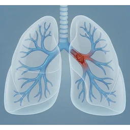
Pulmonary embolism risk stratification using clinical parameters to estimate mortality and guide management decisions

Coronary artery disease reporting on CT angiography using standardized stenosis severity and reporting categories.

Lung cancer screening reporting on CT using standardized Lung-RADS categories and recommendations.

Management of incidental pulmonary nodules on CT based on size, morphology, and patient risk factors.

More about the PE-RADS v2026 Calculator
The Pulmonary Embolism Reporting and Data System (PE-RADS) v2026 is a standardized reporting framework for acute pulmonary embolism (PE) evaluated with CT angiography or MR angiography. It combines the anatomic location of the most proximal acute pulmonary embolus with selected image-quality and right-heart findings to support consistent communication in the imaging report.[1]
PE-RADS is intended to accompany a complete interpretation of the examination. The classification does not replace description of clot distribution, image quality, associated findings, or relevant cardiovascular findings.[1]
PE-RADS anatomic categories
When definite acute PE is present, the primary PE-RADS category is determined by the most proximal acute embolus identified in the pulmonary arterial tree.[1]
| PE-RADS | Most proximal acute PE finding |
|---|---|
| 0 | No acute PE |
| 1 | Subsegmental pulmonary artery |
| 2 | Segmental pulmonary artery |
| 3 | Lobar or interlobar pulmonary artery, past the takeoff of the upper-lobe artery |
| 4 | Central pulmonary artery: main, right, or left pulmonary artery |
The score is assigned on a per-patient basis. If emboli are present at several levels, the most proximal acute embolus determines the ordinal category. PE-RADS 3 includes a lobar artery and vessels past the upper-lobe artery takeoff, including the interlobar artery and basal trunk to the lower lobe.[1]
Limited and nondiagnostic examinations
PE-RADS distinguishes a limited examination from a nondiagnostic examination when no definite acute PE is identified.[1]
PE-RADS 0L is used when no acute PE is identified but assessment is limited at the segmental and/or subsegmental pulmonary arterial level. The report should document the level to which the pulmonary arteries are confidently assessed. Clinical judgment then determines whether further evaluation is needed.[1]
PE-RADS N is used when image quality is inadequate to reliably exclude acute PE in the main and/or lobar pulmonary arteries. A PE-RADS N examination cannot be used to guide patient management, and further evaluation is required. The PE-RADS management table recommends consideration of alternative imaging or repeat CT/MR angiography.[1][2]
The L and N designations should not replace an ordinal category when a definite acute pulmonary embolus has already been identified.[1]
PE-RADS E exception modifier
The E modifier identifies an intravascular pulmonary arterial filling defect that does not represent acute pulmonary thromboembolism.[1]
Examples include chronic thromboembolism, in situ thrombus, pulmonary artery sarcoma, tumor embolus, cement or other exogenous-material embolus, and air embolus. This list is not comprehensive. When E is used, the report should describe both the location and presumptive type of the non-acute filling defect in the Findings and Impression.[1]
The defining publication gives examples such as PE-RADS 0/E when a non-acute pulmonary arterial filling defect is present without acute PE and PE-RADS 3/E when acute lobar or interlobar PE coexists with a separate non-acute filling defect.[1]
Right ventricular enlargement: RV+
The RV+ modifier indicates right ventricular enlargement defined by an RV-to-LV ratio of at least 1.0.[1]
The RV/LV ratio may be measured on axial images, on a reconstructed four-chamber view, or volumetrically. PE-RADS does not designate one of these measurement methods as superior for outcome prediction.[1]
The framework uses the term RV enlargement rather than RV strain. CT or MR angiography, particularly when prior imaging is unavailable, may not reliably establish whether right ventricular enlargement is acute or chronic.[1]
For patients with acute PE, the clinical significance attributed to RV enlargement varies with clot location. PE-RADS states that acute PE is unlikely to be the sole cause of RV enlargement with PE-RADS 1, while acute central PE in PE-RADS 4 is likely to be a major contributing cause.[1]
Right-heart thrombus: T+
The T+ modifier is used when definite thrombus is identified in the right atrium and/or right ventricle.[1]
Absence of T+ does not mean that right-heart thrombus has been definitively excluded. Variable right-heart enhancement, trabeculation, and cardiac motion may limit evaluation of the right-sided cardiac chambers on PE CT or MR angiography.[1]
Definite right-heart thrombus is clinically important and is incorporated into PE-RADS because it can affect the next step in patient care.[1]
Reporting the PE-RADS classification
The PE-RADS classification combines the primary category with applicable exception and cardiac modifiers. The preferred reporting order is the primary ordinal or nondiagnostic classification, exception when applicable, RV status, and right-heart thrombus status.[2]
Valid illustrative combinations include PE-RADS 4/RV+, PE-RADS 4/RV+/T+, PE-RADS 0/E, and PE-RADS 3/E. The ACR assessment-category note also permits RV+ and T+ descriptors with PE-RADS N and 0L when those accessory findings are definitively present.[2]
The classification should remain part of a complete CT or MR angiography interpretation rather than functioning as a stand-alone report.[1]
Clinical management considerations
PE-RADS includes population-level management and referral considerations associated with the imaging categories and cardiac findings. These considerations are intended to connect standardized imaging findings with appropriate next-step clinical assessment.[1][2]
They are not patient-specific treatment prescriptions. Imaging findings alone cannot define treatment decisions for acute PE. Clinical status, hemodynamics, bleeding risk, laboratory findings, comorbidities, available resources, and other patient-specific factors must also be considered.[1]
The framework therefore separates standardized imaging classification from the broader clinical risk assessment used to decide anticoagulation, surveillance, cardiovascular assessment, reperfusion therapy, specialist consultation, or other management.[1]
Important limitations and application points
PE-RADS v2026 is designed for acute PE assessment on CT and MR angiography.[1][2]
- The most proximal definite acute embolus determines the primary category.
- Image-quality designations 0L and N are used only when no definite acute PE is identified.
- Non-acute intravascular filling defects are described using E and should have their presumptive type and location documented.
- RV+ indicates RV enlargement using an RV/LV ratio of at least 1.0. It does not by itself establish acute right-heart strain.
- T+ requires definite thrombus in the right atrium and/or right ventricle. Absence of T+ does not prove that right-heart thrombus is absent.
- Suspected septic emboli in the appropriate infectious and parenchymal context are outside the PE-RADS grading framework described in the defining publication.
- PE-RADS v2026 does not specify a separate ordinal category for an equivocal or indeterminate pulmonary arterial filling defect.
- PE-RADS classification does not replace clinical risk stratification or patient-specific treatment decisions.
Frequently Asked Questions (FAQs)
How is the PE-RADS category chosen when pulmonary emboli are present at multiple arterial levels?
Use the most proximal definite acute pulmonary embolus. A more proximal clot determines the primary PE-RADS category even when additional emboli are present distally.[1]
What is the difference between PE-RADS 0L and PE-RADS N?
PE-RADS 0L means no acute PE is identified and the examination is adequate through the lobar pulmonary arteries but limited at the segmental and/or subsegmental levels. Clinical judgment determines whether further evaluation is needed. PE-RADS N means acute PE cannot be reliably excluded at the main and/or lobar pulmonary arterial level; the study cannot be used to guide patient management and further evaluation is required, with alternative imaging or repeat CT/MR angiography listed as next-step considerations.[1][2]
What does the E modifier mean?
E indicates an intravascular pulmonary arterial filling defect that does not represent acute PE. Examples include chronic thromboembolism, in situ thrombus, pulmonary artery sarcoma, tumor embolus, exogenous-material embolus, and air embolus. The presumptive pathology and location should be described in the report.[1]
Does the absence of T+ mean there is no right-heart thrombus?
No. T+ is used only when thrombus is definitively identified in the right atrium and/or right ventricle. The PE-RADS publication notes that not using T+ does not establish that clot is absent from these chambers.[1]
Should suspected septic emboli receive a PE-RADS category?
No. The defining PE-RADS publication excludes suspected septic emboli from grading when intravascular embolic findings occur with parenchymal findings suggesting septic emboli and the clinical context supports infection.[1]
Does PE-RADS determine how a patient with acute PE should be treated?
No. PE-RADS provides standardized imaging classification and population-level management considerations. Individual treatment decisions require integration of imaging with clinical status, hemodynamics, bleeding risk, laboratory data, comorbidities, and other patient-specific factors.[1]
References
- Koweek LM, Agarwal P, Brosnahan SB, et al. Pulmonary Embolism Reporting and Data System (PE-RADS) v2026 for the Diagnosis of Acute Pulmonary Embolism on CT and MR Angiography. Radiology. 2026;320(1):e252721. doi:10.1148/radiol.252721. PMID: 42478953.
- American College of Radiology. PE-RADS v2026 Assessment Categories. Release date: July 2026. Accessed October 22, 2026. https://edge.sitecorecloud.io/americancoldf5f-acrorgf92a-productioncb02-3650/media/ACR/Files/RADS/PE-RADS/PE-RADS-Assessment-Categories.pdf

Radiologist and Nuclear Medicine Specialist
Neuroradiology fellow at Johns Hopkins University
UT Southwestern Medical Center alumnus